
This project was completed as part of the 2026 Hampshire Hospitals Green Team Competition.
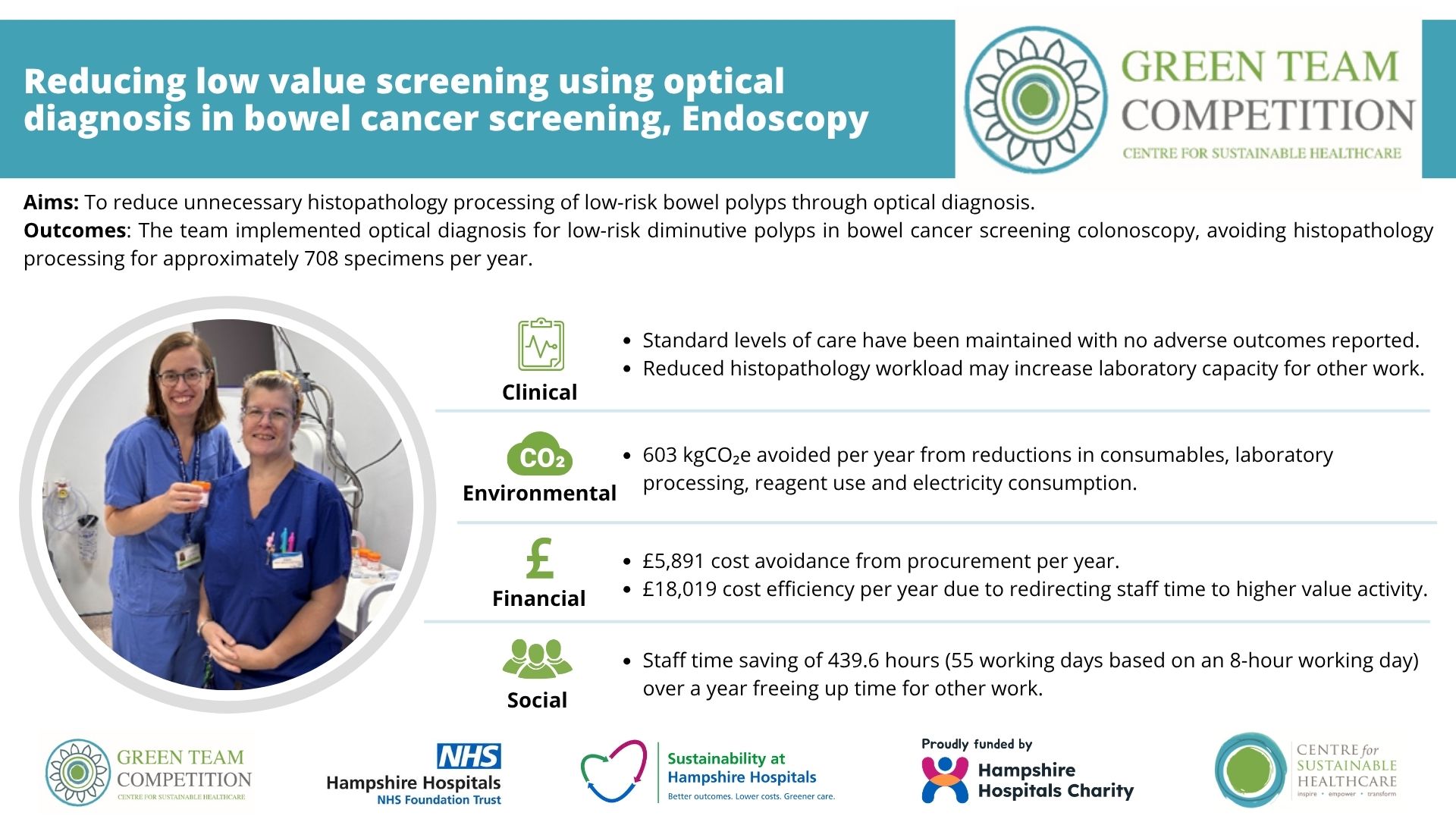
Team members
- Dr Maeve Tierney, Consultant Gastroenterologist
- Kiret Grewal, Senior BMS/Operations Manager, Cellular Pathology
Setting / patient group
This project was undertaken within the bowel cancer screening programme at Royal Hampshire County Hospital. The programme offers screening to adults aged 50-74 years and includes colonoscopy for patients with positive screening results. Historically, all polyps identified during colonoscopy were removed and sent for histopathological assessment.
Issue
Processing bowel polyps through histopathology requires consumables, staff time, energy and laboratory resources, resulting in financial costs and carbon emissions. Recent national guidance introduced optical diagnosis, enabling appropriately trained endoscopists to identify diminutive benign polyps (<5mm) that can be removed and discarded without histopathological analysis. The team sought to quantify the environmental, financial and social benefits associated with this change compared with the previous pathway, where all polyps were sent for histopathology
Intervention
The project evaluated the implementation of optical diagnosis within bowel cancer screening colonoscopy. Data were collected retrospectively from screening lists over a one-month period following implementation. The new pathway allows trained endoscopists to discard low-risk polyps during the procedure, avoiding specimen collection, transport and laboratory processing. Larger polyps or those with concerning features continued to be sent for histopathology in line with national guidance. During the study period, an average of 59 specimens per month, equivalent to 708 specimens annually, avoided histopathology processing
Potential Outcomes
Clinical
The project aimed to maintain existing standards of care while reducing environmental and financial costs. No adverse outcomes were reported following implementation, and safeguards were in place to ensure that any polyps not meeting discard criteria continued to undergo histopathological assessment. Optical diagnosis was implemented as part of the national bowel cancer screening programme, supported by specialist training, competency assessment and photographic documentation of discarded polyps.
Environmental
The assessment found that processing a polyp through histopathology generated approximately852 kgCO₂e. By avoiding the histopathological processing of 708 specimens annually, the project estimated an annual reduction of 603 kgCO₂e. These savings resulted from reductions in consumables, laboratory processing, reagent use and electricity consumption throughout the pathology pathway.
Financial
The project identified savings from reduced use of consumables and staff time. Procurement savings were estimated at £5,891 per year, including reductions in polyp traps, gloves and slide processing. Staff time savings were estimated at £18,019 per year, based on avoiding at least 37.25 minutes of processing time per specimen. Combined annual savings were estimated at £23,909.
Social
Optical diagnosis was estimated to save approximately 439.6 staff hours per year, equivalent to around 55 working days. Reduced workload within endoscopy and histopathology enables skilled staff to focus on other activities and increases laboratory capacity for higher-priority work. Staff were reported to be highly supportive of the initiative and recognised its environmental, financial and social benefits. The project also highlighted the potential for more efficient use of healthcare resources while maintaining patient care standards.
Key learning point
The project demonstrated that significant environmental, financial and social benefits can be achieved through relatively modest reductions in specimens sent for histopathology. Key learning included the importance of collaborative working between endoscopy and laboratory teams, alongside clear training and guidance for endoscopists undertaking optical diagnosis. The team found that understanding the full laboratory pathway helped identify opportunities for improvement and supported implementation of a change that aligns with national policy while maintaining high standards of patient care.
 Download resource -
Please log in or sign up to comment.